
Definition
Fibrous dysplasia of the ribs is a benign bone condition characterized by the replacement of normal bone tissue with fibrous tissue, causing weakening and increased fragility of the affected ribs and making them more susceptible to fractures. This condition can affect one or more ribs and may sometimes involve other bones, such as the skull or long bones.
Epidemiology
This condition is considered rare, with an estimated prevalence of between 0.5% and 1% of bone diseases. It most commonly affects children and young adults, with an average age of onset between 10 and 30 years. However, it can occur at any age.
Symptoms
Symptoms can vary considerably. Some individuals may be completely asymptomatic, while others may experience localized pain on one side of the chest, deformities or bony prominences in the rib area and, in more severe cases, breathing difficulties due to compression of surrounding structures.
Diagnosis
Diagnosis is based on a thorough physical examination combined with various diagnostic methods, including radiographic examinations that may show characteristic bone lesions and areas of deformity.
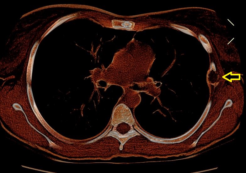
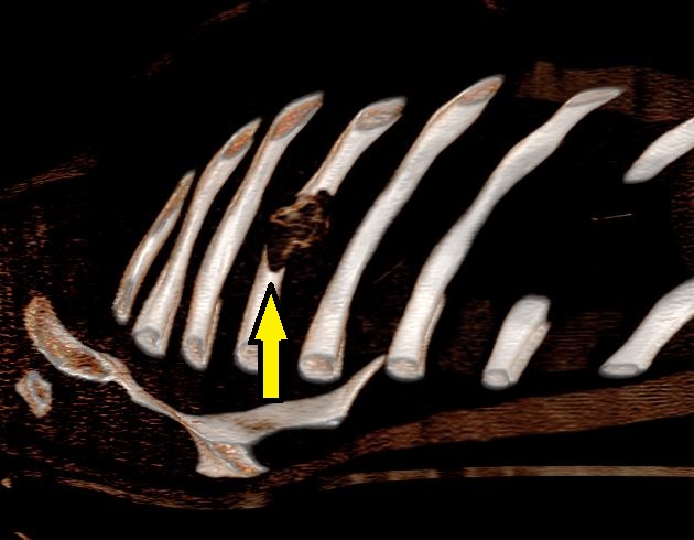
Chest computed tomography (CT) is fundamental in this context (Figure 1), also with the use of 3D reconstructions (Figure 2), which can provide a detailed assessment of the structure of the affected bone. Magnetic resonance imaging (MRI) can be helpful in cases requiring differential diagnosis.
Bone biopsy is strongly recommended, especially when imaging findings are unclear and there is suspicion of chondrosarcoma or another more aggressive bone tumor. Biopsies are performed under local anesthesia by Diagnostic and Interventional Radiology and can confirm the presence of abnormal fibrous tissue within the affected bone.
Treatment
Treatment depends on the severity and location of the dysplasia. In mild cases, regular monitoring, for example with CT and/or MRI examinations every six months, and pain management with analgesics may be indicated.
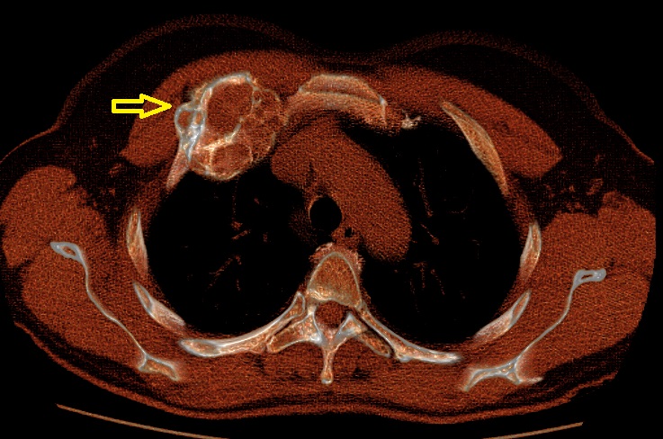
However, in more severe cases causing significant symptoms or deformity (Figure 3), surgery may be necessary to remove the affected bone segment or segments and stabilize the remaining bone structures, reducing pain and improving function.
Monitoring and Follow-up
After treatment, regular follow-up is essential to assess the progression of the disease or, in the case of surgery, the postoperative outcome. This involves periodic imaging examinations and, if necessary, assessment of respiratory function to monitor any long-term complications.
Author: Dr. Fabio Davoli, Head of the General and Thoracic Surgery Unit, Rizzoli Orthopedic Institute.
Scheda revisionata il: 12 dicembre 2023