
Elastofibroma dorsi is a rare benign soft-tissue tumor composed predominantly of elastic and fibrous connective tissue. It typically develops in the subscapular region, between the scapula and the chest wall, and is often asymptomatic. Although the exact cause of elastofibroma dorsi is not fully understood, its development appears to be associated with repetitive mechanical stress involving the shoulder girdle, chronic microtrauma, and prolonged friction or pressure between the scapula and the chest wall. The condition is frequently bilateral.
Diagnostic Tests
The diagnosis of elastofibroma dorsi is based on a combination of clinical examination and imaging studies, with biopsy required only in selected cases. On physical examination, the physician may identify a palpable, usually non-tender mass in the subscapular region, which may become more prominent during forward movement of the shoulder and scapula. Imaging studies, particularly Computed Tomography (CT) and Magnetic Resonance Imaging (MRI) (Figure 1),
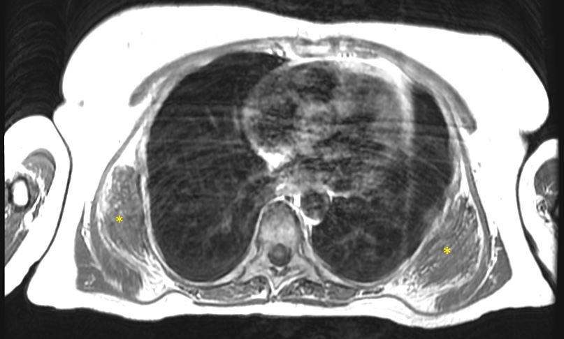
are used to confirm the presence of the lesion, assess its size and anatomical relationships, and evaluate the surrounding tissues. Biopsy is not routinely required when clinical and radiological findings are typical. However, tissue sampling may be indicated when imaging findings are atypical or raise suspicion of a malignant soft-tissue tumor.
Treatment
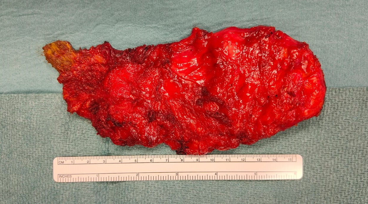
In many cases, elastofibroma dorsi does not require active treatment, particularly when the lesion is small, asymptomatic, and does not cause functional impairment. Surgical treatment may be considered when the lesion becomes large or symptomatic, causes significant discomfort or pain, or interferes with normal shoulder and scapular movement. Surgery is performed under general anesthesia and consists of complete excision of the lesion through a subscapular incision, with careful preservation of the surrounding muscles and other anatomical structures (Figure 2).
Post-operative course
The usual post-operative hospital stay is approximately 4-5 days, with particular attention paid to pain management and wound care. Potential complications include bleeding, surgical-site infection, and post-operative seroma formation. Bleeding and infection are relatively uncommon, whereas seroma formation is more frequent, particularly after the removal of large lesions. When clinically indicated, a post-operative seroma may require one or more percutaneous aspirations. In selected cases, microbiological analysis of the aspirated fluid and antibiotic therapy may also be necessary.
Patients may occasionally be discharged with a subfascial drain in place. The drain can subsequently be removed once the daily volume of fluid has decreased to an appropriate level. Patients are generally followed closely during the first month after surgery. A further surgical follow-up visit is recommended approximately three months after the procedure, usually accompanied by an ultrasound examination of the soft tissues in the scapular region.
In conclusion, elastofibroma dorsi is a rare benign soft-tissue tumor that requires an individualized approach to management. Accurate diagnosis based primarily on clinical assessment and imaging allows patients with typical asymptomatic lesions to be managed conservatively, while surgical excision can provide effective treatment for symptomatic or functionally limiting lesions.
Author: Dr. Fabio Davoli, head of General & Thoracic Surgery Unit, IRCCS Rizzoli Orthopaedic Institute.
Scheda revisionata il: 13 novembre 2023.