
Rib exostoses are abnormal bony growths that develop on the surface of the ribs in the thoracic region. These bony formations can vary in size and may be single or multiple. Although they are often asymptomatic, in some cases they can cause pain, compression of surrounding structures, or visible deformities resulting in cosmetic concerns. The most concerning complication is the possible, although rare, malignant transformation of these lesions.
Epidemiology – Associated Diseases
Rib exostoses are considered rare, and their incidence varies. They are more commonly observed in males and generally become apparent during adulthood, although they can also occur in the pediatric population.
The exact cause of rib exostosis formation is not completely understood, but it is believed that genetic and environmental factors may play a role. They can be associated with conditions such as Hereditary Multiple Exostoses (HME), a genetic disorder characterized by the development of multiple exostoses in different parts of the body, generally involving the limbs and scapulae (with a prevalence of approximately 1 in 50,000 births). In addition, they may be associated with disorders of bone development.
Diagnosis
The diagnosis of rib exostoses includes a thorough physical examination, which may detect the presence of bony protrusions or growths arising from the ribs. Imaging examinations, such as X-rays, computed tomography (CT), or magnetic resonance imaging (MRI), are essential to evaluate the size of the exostoses, their relationship with surrounding structures, and to rule out other similar conditions. In pediatric patients, there is a tendency to favor the use of MRI.
Biopsy is rarely necessary but may be performed to confirm the benign nature of the bone lesion. In general, it is preferable to obtain the definitive histological diagnosis after the lesion has been surgically removed.
Treatment
The treatment of rib exostoses depends on the severity of symptoms, their size, and their radiological characteristics. Typically, pediatric patients are observed until the age of 13–14 years, with early surgical intervention reserved for large exostoses that compromise limb function, for example in the case of large exostoses involving the shoulder girdle.
In adult patients, cosmetic concerns may represent another indication for treatment. If the radiological characteristics raise suspicion of transformation into a chondrosarcoma, surgical excision is also indicated.
In the case of asymptomatic exostoses, management can be conservative, with periodic monitoring and pain management if necessary. The objective of surgery is to remove the exostosis without compromising the structure of the rib and while minimizing the risk of recurrence.
Surgical Techniques
From a technical point of view, surgical removal of a rib exostosis can be performed:
- extra-thoracically (Figure 1);
- through an intrathoracic approach, accessing the pleural cavity by thoracotomy;
- through a minimally invasive intrathoracic approach, accessing the pleural cavity using video-assisted thoracic surgery (VATS) (Figure 2).
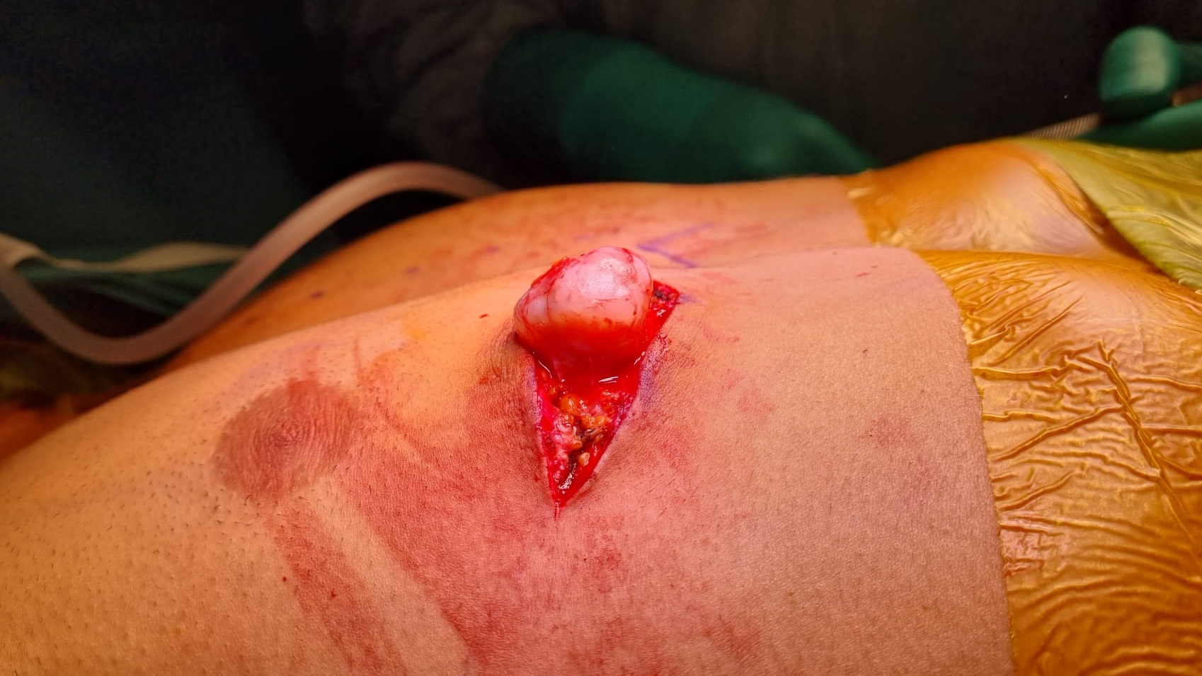
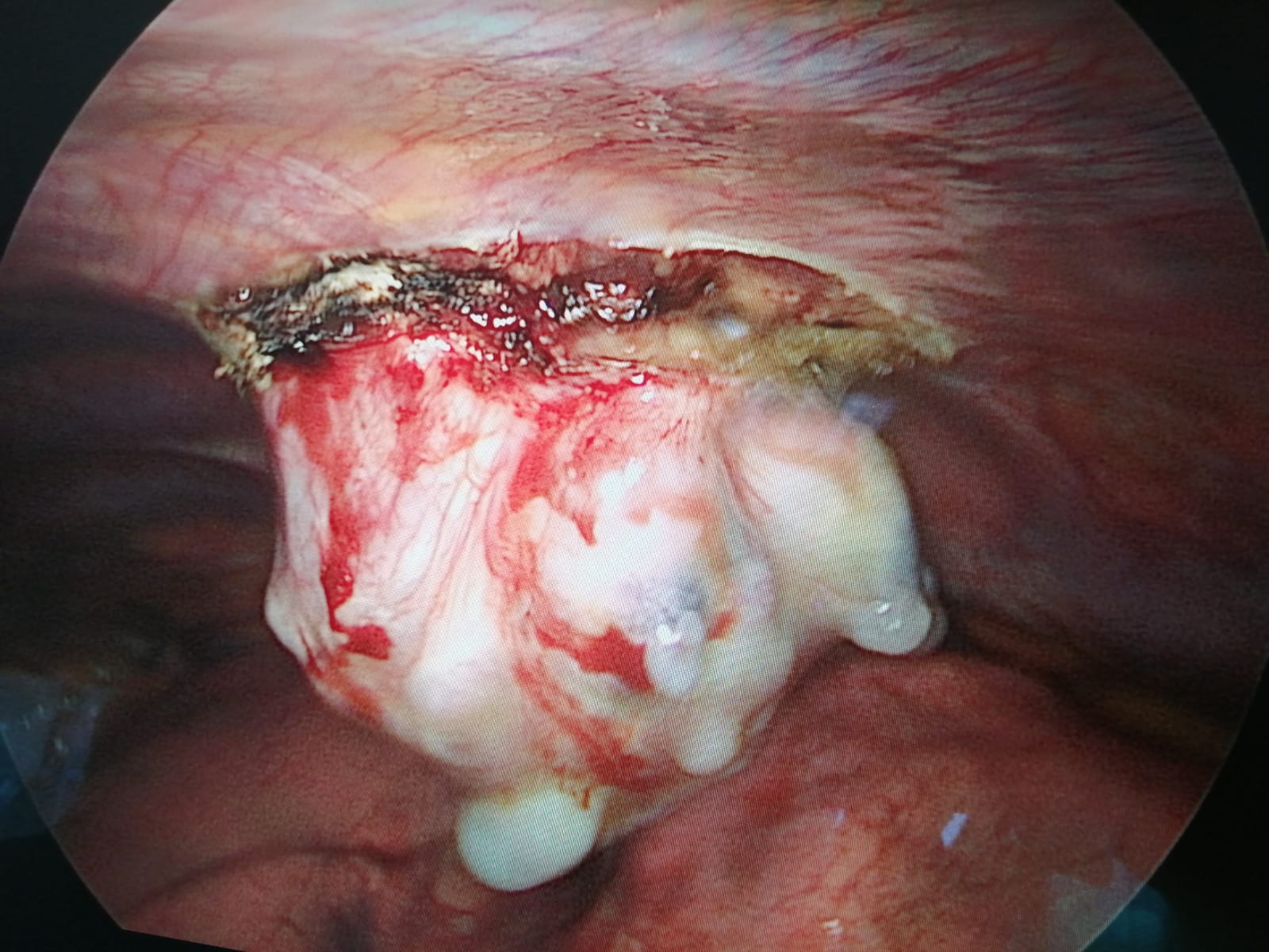
Regardless of the technique used, the objective is to remove the exostosis while preserving the involved rib and chest wall structures as much as possible. In some cases, it may be necessary to use a Gore-Tex prosthesis to cover the resulting chest wall defect.
In summary, rib exostoses are abnormal bone formations that require careful evaluation and personalized management based on symptoms and their impact on the patient's quality of life. Early diagnosis and a targeted therapeutic approach can help reduce symptoms and improve quality of life.
Author: Dr. Fabio Davoli, Head of the General and Thoracic Surgery Unit, Rizzoli Orthopedic Institute.
Scheda revisionata il: 15 novembre 2023.