
Lipomas are benign tumors made up of fatty tissue. In the chest, they most commonly develop within the subcutaneous tissues of the chest wall, although they may occasionally arise deeper, between or within muscle layers.
They are among the most common benign soft-tissue tumors. Lipomas usually grow slowly, are soft and mobile, and are generally painless. Although they are usually harmless, they may cause cosmetic concerns, discomfort or, particularly when they become large or develop in deeper locations, compression of nearby structures and functional problems.
Diagnosis
The diagnosis of a chest lipoma usually begins with a clinical examination. The doctor may identify a soft, mobile and generally painless mass beneath the skin.
Imaging examinations, such as ultrasound or magnetic resonance imaging (MRI), may be used to confirm the fatty nature of the mass, assess its size and location, and evaluate its relationship with surrounding structures, particularly muscles and the deeper tissues of the chest wall.
In selected cases, especially when imaging shows atypical features or when the mass is large, deep or rapidly growing, a biopsy may be required to distinguish a simple lipoma from an atypical lipomatous tumor or other soft-tissue tumors.
Treatment
Small, asymptomatic lipomas with typical clinical and imaging features do not always require surgical treatment. Depending on their characteristics, clinical observation and periodic imaging may be appropriate.
Surgical removal may be recommended when a lipoma causes pain or discomfort, increases in size, interferes with daily activities, causes a significant cosmetic problem, or when its clinical or imaging characteristics do not allow a confident diagnosis.
Surgery is also frequently considered for particularly large or deep lipomatous masses (Figure 1),
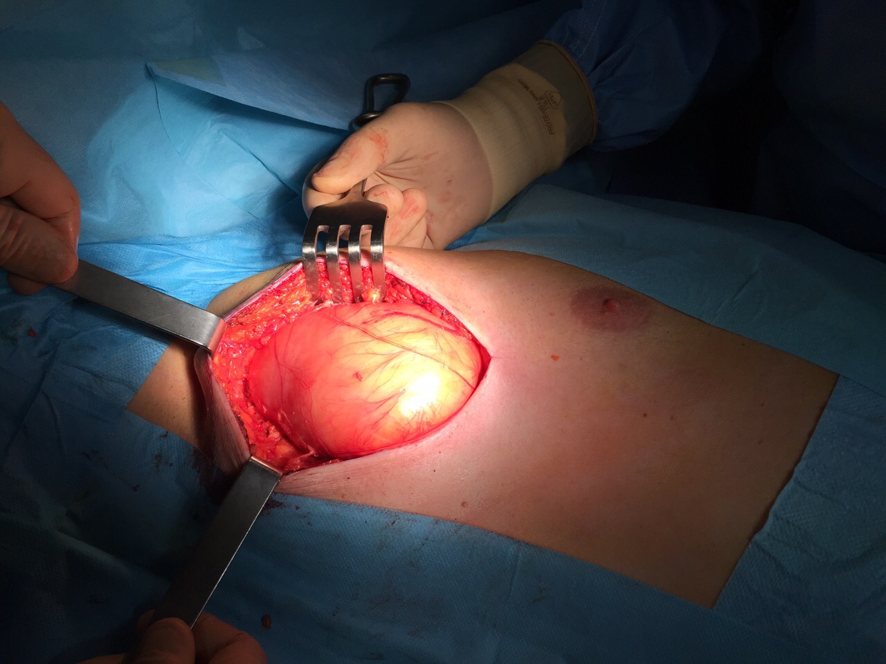
for which complete assessment is important to exclude an atypical lipomatous tumor or other diagnoses.
During surgery, the aim is to remove the mass completely while preserving the function and appearance of the surrounding chest wall as much as possible. The risk of recurrence after complete removal of a simple lipoma is generally low.
Depending on the size, depth and location of the mass, surgery may be performed under local or general anesthesia. Hospital stay is usually short and, in selected cases, the procedure can be performed on a day-surgery basis.
Overall, chest lipomas are benign tumors composed predominantly of fatty tissue. Clinical assessment and appropriate imaging usually allow an accurate diagnosis, while surgery provides definitive treatment when indicated. Management should always be tailored to the characteristics of the individual patient and the specific lesion.
What Are Atypical Lipomatous Tumors of the Chest?
Atypical lipomatous tumors (ALTs) are fatty tumors that differ from simple lipomas in their biological behavior and microscopic characteristics.
Compared with simple lipomas, they may be larger, deeper and more closely associated with muscles and other structures of the chest wall. On imaging, they may also have a more complex or irregular appearance (Figure 2).
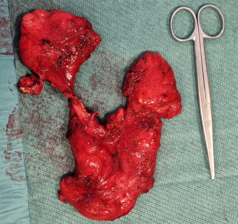
When an atypical lipomatous tumor is suspected, MRI is particularly useful for defining its extent and relationship with the surrounding anatomical structures. A biopsy is generally required before surgical treatment in order to establish the diagnosis and plan the most appropriate surgical approach.
The management of atypical lipomatous tumors may require a multidisciplinary approach involving surgeons, radiologists, pathologists and oncologists. Accurate assessment of their anatomical characteristics and biological behavior is important in determining the optimal treatment strategy.
Surgical excision is generally the treatment of choice for atypical lipomatous tumors of the chest wall. The objective is complete removal of the tumor while preserving, whenever possible, the function and stability of the chest wall.
Unlike simple lipomas, atypical lipomatous tumors have a greater tendency to recur locally after surgery. In addition, although their metastatic potential is very low in the absence of dedifferentiation, long-term follow-up is important because these tumors may occasionally undergo dedifferentiation into a more aggressive form.
After surgical excision, regular follow-up is therefore recommended. The type and frequency of imaging examinations will depend on the location and characteristics of the tumor, the surgical margins and the definitive histological findings, and may be determined within a multidisciplinary setting.
Author: Dr. Fabio Davoli, Head of the General and Thoracic Surgery Unit, Rizzoli Orthopedic Institute.
Scheda revisionata il: 13 novembre 2023.